총간관
Common hepatic duct| 총간관 | |
|---|---|
 | |
| 세부 사항 | |
| 식별자 | |
| 라틴어 | 간관 |
| 메쉬 | D006500 |
| TA98 | A05.8.01.061 |
| TA2 | 3092 |
| FMA | 14668 |
| 해부학 용어 | |

총간관은 담도의 첫 번째 부분입니다.그것은 담낭에서 나오는 낭포관과 결합하여 총담관을 형성한다.
구조.
총간관은 [2]담도의 첫 번째 부분입니다.그것은 오른쪽 간관(간의 오른쪽 기능성 엽에서 담즙을 배출하는)과 왼쪽 간관(간의 [3]왼쪽 기능 엽에서 담즙을 배출하는)의 수렴에 의해 형성된다.그런 다음 담낭에서 나오는 낭포관과 결합하여 총담관을 형성합니다.
덕트의 길이는 [4]보통 6~8cm입니다.일반적인 간관은 성인의 경우 직경이 약 6mm이며 약간의 변화가 [4]있다.내부 표면은 단순한 주상피로 [3]덮여있다.
변화
약 1.7%의 사람들이 공통 [5]간관에 연결되는 추가적인 부속 간관을 가지고 있다.
간관이 담낭에 직접 결합하는 경우는 드물기 때문에 [5]질병으로 이어집니다.
기능.
간관은 간에서 장으로 분비물을 운반하는 담도의 일부이다.
임상적 의의
담낭 절제술
일반적인 간관은 [citation needed]담낭을 제거한 사람들에게 더 많은 양의 담즙을 운반한다.
총간관은 담낭절제술과 같은 수술에서 중요한 해부학적 표식이다.그것은 낭포관, 낭포동맥과 함께 칼로 삼각형의 한쪽 끝을 형성한다.이 삼각형의 모든 구성요소는 잘못된 구조를 자르거나 자르지 않도록 식별해야 합니다.
콜레스트시스
지름이 8mm 이상인 경우는 비정상적인 팽창으로 간주되며, 담즙의 [6]증세를 나타냅니다.
미리치 증후군
미리지 증후군은 담석으로 [7]인해 간관이 막혀 버렸을 때 발생합니다.
기타 이미지
이 해부학적 특징 갤러리는 스타일의 의료 매뉴얼을 준수하기 위해 청소가 필요합니다. |
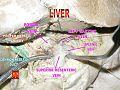
총간관

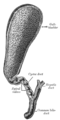
간문맥과 그 지류.
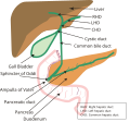
담낭과 담관이 열려 있었다.

총간관


레퍼런스
- ^ Standring S, Borley NR, eds. (2008). Gray's anatomy : the anatomical basis of clinical practice. Brown JL, Moore LA (40th ed.). London: Churchill Livingstone. pp. 1163, 1177, 1185–6. ISBN 978-0-8089-2371-8.
- ^ Manohar, Rohan; Lagasse, Eric (2014-01-01), Lanza, Robert; Langer, Robert; Vacanti, Joseph (eds.), "Chapter 45 - Liver Stem Cells", Principles of Tissue Engineering (Fourth Edition), Boston: Academic Press, pp. 935–950, doi:10.1016/b978-0-12-398358-9.00045-8, ISBN 978-0-12-398358-9, retrieved 2021-01-26
- ^ a b Bergman, Simon; Geisinger, Kim R. (2008-01-01), Bibbo, Marluce; Wilbur, David (eds.), "CHAPTER 14 - Alimentary Tract (Esophagus, Stomach, Small Intestine, Colon, Rectum, Anus, Biliary Tract)", Comprehensive Cytopathology (Third Edition), Edinburgh: W.B. Saunders, pp. 373–408, ISBN 978-1-4160-4208-2, retrieved 2021-01-26
- ^ a b 그레이 아나토미, 제39판, 1228페이지
- ^ a b Portmann, Bernard C.; Roberts, Eve A. (2012-01-01), Burt, Alastair D.; Portmann, Bernard C.; Ferrell, Linda D. (eds.), "3 - Developmental abnormalities and liver disease in childhood", MacSween's Pathology of the Liver (Sixth Edition), Edinburgh: Churchill Livingstone, pp. 101–156, ISBN 978-0-7020-3398-8, retrieved 2021-01-26
- ^ Hoeffel, Christine; Azizi, Louisa; Lewin, Maité; Laurent, Valérie; Aubé, Christophe; Arrivé, Lionel; Tubiana, Jean-Michel (2006). "Normal and Pathologic Features of the Postoperative Biliary Tract at 3D MR Cholangiopancreatography and MR Imaging". RadioGraphics. 26 (6): 1603–1620. doi:10.1148/rg.266055730. ISSN 0271-5333. PMID 17102039.
- ^ Katz, Seth S. (2017-01-01), Jarnagin, William R. (ed.), "Chapter 18 - Computed tomography of the liver, biliary tract, and pancreas", Blumgart's Surgery of the Liver, Biliary Tract and Pancreas, 2-Volume Set (Sixth Edition), Philadelphia: Elsevier, pp. 316–357.e6, ISBN 978-0-323-34062-5, retrieved 2021-01-26
외부 링크
- 해부사진: 38:03-0302 SUNY 다운스테이트 메디컬센터 - "위, 비장, 간: 간십이지장 인대 내용물"
- 일러스트


