세르톨리 세포종양
Sertoli cell tumour| 세르톨리 세포종양 | |
|---|---|
 | |
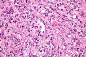
| 세르톨리 세포 종양의 마이크로그래프. H&E 얼룩. | |
| 전문 | 종양학 |
세르톨리 세포 종양, 또한 세르톨리 세포 종양(미국 철자법)은 세르톨리 세포의 성 코드-고나달성 척추 종양이다. 그것들은 고환이나 난소에서 발생할 수 있다. 그것들은 매우 희귀하며 일반적으로 35세에서 50세 사이에 최고조에 이른다. 그것들은 전형적으로 잘 구분되어 있으며, 매우 유사하게 나타나는 경우가 많기 때문에 세미노마로 오진될 수 있다.[1]
세르톨리 세포와 레이디그 세포를 모두 생성하는 종양은 세르톨리-라이디그 세포 종양으로 알려져 있다.
프리젠테이션
남성의 경우 Sertoli 세포 종양은 전형적으로 고환질량 또는 단단함으로 나타나며, 특히 안드로겐을 생산하는 경우에는 어린 소년들에게서 성조숙증(25%)을 동반할 수 있다.[2]
진단
초음파에서 Sertoli 세포 종양은 보통 혼자 있는 저자극성 체내 병변으로 나타난다. 그러나 큰 셀 하위 유형은 석회화 영역이 큰 다중 질량 및 양자 질량으로 나타날 수 있다. MRI도 시행될 수 있지만, 이것은 전형적으로 확정적이지 않다.[2]
현미경 검사와 면역 화학은 특히 반종이 의심될 때 결정적인 진단을 내릴 수 있는 유일한 방법이다.[3]
치료
남성의 경우 영상기법으로 종양을 식별하기 어려워 난초절제술을 하는 경우가 많다. 세르톨리 세포 종양의 대다수는 양성이다. 따라서 이 정도면 충분하다. 화학요법이나 방사선 치료의 문서화된 이점은 없다.[4]
비인간에서
세르톨리 세포 종양은 국내산 오리,[5] 개,[6][7] 말을 포함한 다른 종에서 발생하는 것으로 알려져 있다.
추가 이미지
레이디그 세포 종양의 마이크로그래프.
참고 항목
메모들
- ^ Young, Robert H. (2005-01-01). "Sex cord-stromal tumors of the ovary and testis: their similarities and differences with consideration of selected problems". Modern Pathology. 18 (S2): S81–S98. doi:10.1038/modpathol.3800311. ISSN 0893-3952. PMID 15502809.
- ^ a b Morgan, Matt A. "Sertoli cell tumour of the testis Radiology Reference Article Radiopaedia.org". radiopaedia.org. Retrieved 2016-12-06.
- ^ Henley, John D.; Young, Robert H.; Ulbright, Thomas M. (2002-05-01). "Malignant Sertoli cell tumors of the testis: a study of 13 examples of a neoplasm frequently misinterpreted as seminoma". The American Journal of Surgical Pathology. 26 (5): 541–550. doi:10.1097/00000478-200205000-00001. ISSN 0147-5185. PMID 11979085. S2CID 33939867.
- ^ "Testis and epididymis - Sertoli cell tumor, NOS". www.pathologyoutlines.com. Retrieved 2016-12-06.
- ^ Leach S, Heatley JJ, Pool RR, Spaulding K (December 2008). "Bilateral testicular germ cell-sex cord-stromal tumor in a pekin duck (Anas platyrhynchos domesticus)". J. Avian Med. Surg. 22 (4): 315–9. doi:10.1647/2007-017.1. PMID 19216259. S2CID 24196361.
- ^ Gopinath D, Draffan D, Philbey AW, Bell R (December 2008). "Use of intralesional oestradiol concentration to identify a functional pulmonary metastasis of canine sertoli cell tumour". J Small Anim Pract. 50 (4): 198–200. doi:10.1111/j.1748-5827.2008.00671.x. PMID 19037884.
- ^ Vegter AR, Kooistra HS, van Sluijs FJ, van Bruggen LW, Ijzer J, Zijlstra C, Okkens AC (October 2008). "Persistent Mullerian Duct Syndrome in a Miniature Schnauzer Dog with Signs of Feminization and a Sertoli Cell Tumour". Reprod. Domest. Anim. 45 (3): 447–52. doi:10.1111/j.1439-0531.2008.01223.x. PMID 18954385.
외부 링크
 위키미디어 커먼스의 세르톨리 세포종양 관련 매체


